


-
Foot-and-mouth disease (FMD) is a highly con-tagious disease of cloven hoofed animals including domesticated ruminants and pigs (14, 17, 22). Once outbreaks of FMD occur, economics losses can be huge. For example, in 1997, an outbreak of FMD occurred in the Taiwan province of China, and total losses, including direct and additional losses to the economy, were estimated to be US$ 1.9 billion (3, 19). Previous reports indicated that the virus resides in the bodily fluids and secretions of infected animals such as the vesicular epithelium and fluid, saliva, milk, faeces, urine, semen and vaginal secretion. Therefore it is very important to detect possible infections and origins of contamination in order to prevent and control FMD and reduce subsequent impact.
Nowadays, the epidemiology of animal viral diseases has been changed significantly by intro-duction of artificial insemination. Many economically important animal viruses have been detected and isolated from boar semen (8, 9, 15, 20, 21). In December 2005, 48 out of 91 seed bulls in a bull farm in Shandong province were found to be showing clinical signs of FMD. To investigate the possibility that bull semen was contaminated with FMDV, 15 bull semen samples including 5 samples from bulls with clinical signs of FMD were collected and analysed by RT-PCR and virus isolation.
HTML
-
The fifteen bull semen samples were collected from a bull farm in Shandong province, 5 of which exhibited typical clinical signs of FMD. The remaining 10 animals appeared normal. All samples were stored at -80℃ until use.
-
Virus RNA was extracted from bull semen samples using RNAiso Reagent (Takara) according to the manufacture's instructions. 5 μL of the extracted RNA was added into the reaction mixture (25 μL) which contained 2.5μL of 10×one step RNA PCR buffer, 500 μmol/mL MgCl2, 200 μmol/mL dNTPs, 40 U RNase in-hibitor, 5 U AMV RTase XL, 5 U AMV-Optimized Taq and 50 pmol/mL of each primer. The first strand cDNA was synthesized at 50 ℃ for 30 min, followed at 94 ℃ for 5 min. PCR amplification was carried out as follows, 94 ℃ for 1 min, 56 ℃ for 30 sec, 72 ℃ for 30 sec, the whole reaction was run for 30 cycles, followed by a final extension of 72 ℃ for 8 min. PCR products were analyzed by 1.2% agarose gel electrop-horesis with ethidium bromide.
-
The VP1 gene of FMDV was purified and sequenced with a CEQTM8000 Genetic Analysis System (Bechman Coulter). The sequencing primers were the sense strand primer 5′-GACTCGAACGTGT CCCTGCCAACT-3′ and anti-sense strand primer 5′-TGCGGTACGGCCACCGTACTACTTC-3′, respec-tively. Alignment and analysis of the nucleotide acid and the amino acid were peformed by using Clustal W method (DNASTAR, USA).
-
The infectivity of 15 semen samples was deter-mined with a monolayer of primary calf thyroid cells. Briefly, 3-day-old monolayer of primary calf thyroid cells were inoculated with 200μL of diluted semen per well in 6-well cell culture plate. The cytopathic effect (CPE) was observed within 48 h post-inoculation. If no CPE was observed in the inoculation cells, the cell culture was stopped and was frozen at -80 ℃ for serial passage. All samples were serially passaged until the third generation. The CPE positive samples were serially passaged for 10 generations.
Semen samples
RNA extraction and RT-PCR
Sequencing and analysis of nucleic acid
Isolation of virus
-
As shown in Fig 1, 4 out of 5 semen samples collected from bulls exhibiting signs of FMD also showed positive results for FMDV RNA by RT-PCR. No amplification products were detected in the others (data no shown). The VP1 gene of FMDV RNA was sequenced and submitted to GenBank on 8 February 2007 (accession number EF428440). Two nucleotide acid sequences from vesicular fluid and semen were aligned by the Clustal W method. The results indicated that virus strain from semen has a close genetic relationship with reference virus strain Asia1/ Jiangsu/ China/2005 (GenBank, accession number DQ-156527), and sequence similarity was up to 97.9% belonging to the same gene sub-group (data no shown). The partial coding sequence at the 3-end of the VP1 gene was analysed by DNASTAR. The results showed that amino acid substitutions were located at five sites: 140(S→T), 141(S→T), 154 (N→S), 176 (I→L) and 211(W→L), and except for two residues at 140 and 141 which were located close to the N terminus of the RGD triplet, the others were located far away from the C terminus of the RGD motif. However the RGD motif was still conserved (Fig. 2).

Figure 1. Analysis of VP1 gene of FMDV by RT-PCR.M, DNA ladder; 1-4, Positive band; 5, Negative.
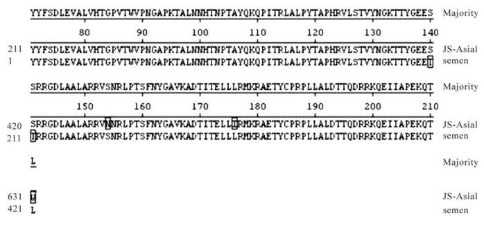
Figure 2. The analysis of partial amino acid sequence of VP1 of FMDV by Clustal W method.
-
No cytopathic effect (CPE) was visualized in all inoculated cells and negative control in the first passage within 48 h post-inoculation. After the second incubation period of 40-48 h, the CPE was observed in the primary calf thyroid cells inoculated semen samples which were positive via RT-PCR (Fig. 3). No CPE was observed in the control cells and other samples. During the serial passages (6-10 passages), the CPE of virus isolated from semen samples was viewed 24 h post-inoculation and the CPE was up to 75% within 36 to 40 h post-inoculation.

Figure 3. The result of the semen sample by virus isolation. The CPE were showed in the cells inoculated with semen sample at 44 h postinoculation.
RT-PCR and sequencing
Isolation of virus
-
Previous reports have demonstrated that semen is contaminated by animal viruses, and intra-vaginal infection could be caused by contaminated semen via artificial insemination (4, 12). Furthermore, Bastos et al reported that FMD virus SAT3 was isolated from both semen and sheath wash specimens from a naturally infected African buffalo bulls. The results demons-trated that potential transmission event by mating had occurred in the infected herd (2). In this study, the bio-security of semen samples from an infected bull herd were investigated and evaluated with virus isolation assay and RT-PCR of bull semen. The results showed that the semen was contaminated by FMDV. This is the first identifcation of contaminated semen from naturally infected bulls in China. These results underline the importance of detection and identifi-cation of virus in semen is very important for FMD prevention and control when mating and performing artificial insemination. Analysis of the VP1 gene showed that semen strain had 97.9% identity with reference strain Asia1. Analysis of amino acid sequence found four residues substitution located within the hyper-variable region of the VP1 gene. In particular, the two residues which were located close to the RGD motif seems to represent a crucial mutation. Although, many studies showed that the possibility of viral antigenic change will happen due to crucial residue substitution within the hyper-variable region of VP1 gene. But we can not make a definite conclusion about whether viral antigenicity has been changed or there has been loss of capability of the virus bind to cell receptor integrin ligands. However, previous studies have demonstrated that viral genome mutations in FMD often lead to amino acids changes, particularly within the capsid proteins (1, 16). Such changes have been shown to occur during persistent infection both in cell culture (10, 13) and in vivo (18), and have been linked to changes in antigenicity (11), which could contribute to sub-populations of virus escaping clearance by the immune system.
Taken together, there were two isolates of FMDV which survived in the same bull herd when they were infected naturally. The possible explanations are as follows: Firstly, virus mutation occured in the semen during replication of viral genome due to the viral replicase enzyme's lack of proof-reading activity (7). Secondly, FMDV, like other RNA viruses, is a population containing different viral RNA genomes termed as quasispecies. There are many reports which suggest that FMDV exists as a group of related, but non-identical genomes known as quasispecies (1, 5, 6). In summary, this study offers useful data for FMD prevention and control in the future.

 DownLoad: